Friday 2 August 2013

role of microRNA in cancer

Cancer is one of the most dangerous and the most spread diseases all over the world, although it is not infectious.

A lot of people when they know their illness suffer very sad and mostly lost hope in treatment, but Allah is capable of everything.

 any defect in the content of microRNA affects on the cell which increases the breeding terribly and tumor occurs in the organ component of these cell.

Let's explain how it affects the cell and makes carcinogenic ..
In the beginning, as we know, mRNA produced from the DNA which found on the chromosome , which turns into a specific protein by the translation.

MiRNAs are RNA genes which are transcribed from DNA, but are not translated into protein.

MiRNAs are small noncoding RNAs approximately 18–25 nucleotides in length .
MicroRNAs are produced from either their own genes or from introns.

After their maturity by an enzyme Drosha and DGCR8 in the nucleus they go to the cytoplasm.

These miRNAs stop the translation of  mRNA by compiletary of it , that leads lost the ability of enzymes to convert it to protein.


This change in the content of these proteins may causes the cancer and in some cases it may be his inhibitor

 the cancer can be treated in several ways:

Surgery
radiotherapy  
chemotherapy
Read More

Sunday 21 July 2013

western blotting

western blotting is a widely used analytical technique used to detect specific proteins in the given sample of tissue homogenate or extract.
http://www.western-blot.us/uploads/images/templates/and01/170-3836_view.jpg
It uses gel electrophoresis to separate denatured proteins by the length of the polypeptide. The proteins are then transferred to a membrane (typically nitrocellulose or PVDF), where they are probed (detected) using antibodies specific to the target protein.

It Gives You Information on the:
  •  Size of your Protein
  •  Expression Amount of your Protein
    Sodium dodecyl sulfate polyacrylamide gel electrophoresis
    SDS-PAGE


    Used to separate protein molecules according to their sizes.

 Transfer Proteins to a Membrane (blotting)
-Following electrophoresis, the protein must be transferred from the electrophoresis gel to a membrane. There are a variety of methods that have been used for this process, including diffusion transfer, capillary transfer, heat-accelerated convectional transfer, vacuum blotting transfer and electroelution.

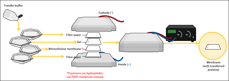
-The transfer method that is most commonly used for proteins is electroelution or electrophoretic transfer because of its speed and transfer efficiency. Electrophoretic transfer of proteins involves placing a protein-containing polyacrylamide gel in direct contact with a piece of nitrocellulose or PVDF membrane and "sandwiching" this between two electrodes submerged in a conducting solution.

-When an electric field is applied, the proteins move out of the polyacrylamide gel and onto the surface of the membrane, where the proteins become tightly attached.

There are two types of apparatus for electrophoretic transfer :
A-wet transfer:
the gel/blotting paper/filter paper sandwich is placed into a cassette along with protective fiber pads. The cassette is then immersed in a buffer tank and subjected to an electrical field. The electric field used for the transfer is oriented perpendicular to the surface of the gel causing proteins to move out of the gel and onto the blotting membrane, which sits between the gel surface and the positive electrode.

B-semi-dry transfer:
the gel/blotting paper/filter paper sandwich is assembled on large electrode
plates which generate the electric field, and buffer is confined to the stack
of wet filter papers.
In semi dry blotting the gel and the membrane are horizontally between two stacks of buffer wetted filter papers in direct contact with two closely spaced solid plate electrodes .the term semi dry refers to the limited amount of buffer that is confined to the stacks of filter papers.

http://upload.wikimedia.org/wikipedia/commons/9/93/Western_blot_transfer.png

-After transfer and before proceeding with the Western blot, total protein on the membrane is often stained with a dye, such as Ponceau S or amido black 10B, to check the transfer efficiency, the gel may also be stained to confirm that protein has been moved out of the gel.
Read More

Friday 19 July 2013

Gel Electrophoresis

Gel electrophoresis is a way to separate and analysis of large molecules, such as proteins, by migrating a colloidal solution of them through a gel. It is also used to sort  DNA strands according to the length.
It is used in a wide range of molecular research and can be used for genotyping or to identify clones that have the correct DNA insert.

http://www.bio.miami.edu/~cmallery/150/gene/DNA_electrophoresis.jpg

How To Use Gel Electrophoresis To Separate DNA

Proteins and nucleic acid molecules can be separated and analysis through gel electrophoresis. it can separate according to the size or charge of these molecules.  the gel which used in electrophoresis is called "agarose gel", and the DNA is separated by an electric current. The gel is located on a small plastic support "an Erlenmeyer flask" along with a salt water buffer. The longer or heavier the DNA fragment, the more slowly it will migrate in the gel. The DNA fragments, produced by PCR , are separated and viewed as bands on the gel. Each gel band contains thousands of DNA molecules of the same size.

http://media-3.web.britannica.com/eb-media/72/47672-004-4E16B61F.jpg


the steps


1. The agarose gel has wells into which the DNA can be loaded by pipette. The gel which located on a small plastic support, is then flooded in a tank filled with liquid buffer and this tank has electrodes on either end. Near the wells is the negative pole, and to the opposite side of the wells is the positive pole.

2. as we know  that DNA has a negative charge, so when there is a current, it will migrate towards the anode ( the positive pole).
 Longer DNA fragments will move through the gel more slowly than the shorter ones. In the following figure, the different DNA bands are shown in blue, but in reality, the DNA is not visible as it moves through the gel so ..

3.there is Post migration step,the gel is removed and the bands gets dyed by ethidium bromide, so the banding patterns of the grouped DNA become visible, and can be analyzed. The ethidium bromide intercalates between DNA bases and causes the DNA fragments to glow when exposed to UV light.

Now the approximate lengths of the unknown DNA strands can be determined by comparing it to the DNA standard.


http://www.tcnj.edu/~elderkin/images/Gallery/Fluoressing%20Gel.jpg
Read More

Monday 15 July 2013

introduction of Gene Cloning


Cloning is a valuable tool in molecular biology because it allows the multiplication of particular genes or proteins that are being studied.

Cloning vectors
Plasmid, Lambda (phage) and cosmid


Features of the useful vector
-Vector can be replicated in a host cell
-Vector and ligated DNA can be replicated in the host cell
-When the vector is in the host cell it gives a novel phenotype or at least easy to be identified


 Basic Skills Needed to Carry Out a simple Gene Cloning Experiment

1- preparation of pure samples of DNA-  amplification

2-Cutting DNA molecules

3-Analysis of DNA fragment sizes

4-Joining DNA molecules together

5-Introduction of DNA into host cells

6-Identification of cells that contain recombinant DNA molecules


 Why the Polymerase Chain Reaction (PCR) is Also Important
  • In a PCR experiment, a single segment of a DNA molecule is copied many times, resulting in an amplified DNA fragment
  • The experiment is designed so that the segment of DNA that is amplified is one that carries the gene of interest
Steps in gene cloning

 the cloning of any DNA fragment:
 (1) select the host organism and cloning vector.
(2) Preparation of vector DNA.
(3) Preparation of DNA to be cloned.(4)Treatment of plasmid and required DNA with the same restriction enzyme
(5) making the recombinant DNA by Mixture of foreign DNA with chopped plasmids and
Addition of DNA ligase.
(6) Introduction of recombinant DNA into host cell.
(7) Production of multiple gene copies & selection process for transformed cells.
(8) examination of clones with required DNA inserts and biological properties.


DNA modifying enzymes

  • Methylation/Acetylation enzymes
  • Nuclease enzymes (ex. endonucleases)
  • Ligation enzymes (Ligase)

Restriction enzyme

Definition
Is a nuclease that cleaves duplex DNA at a particular short sequence (restriction site)
In a symmetrical shape (palindrome)

Mechanism of restricition
By breaking the phosphodiester bond in the DNA backbone creating a free
two 3’ end and 5’ end

Types of restriction products:
Sticky ends producing type
Blunt end producing type

Features:
Cut in a symmetric way
Restrict the DNA regardless its source
Read More

Friday 12 July 2013

siRNA

Small interfering RNA (siRNA)  known as short interfering RNA or silencing RNA, is a class of double-stranded RNA molecules, 20-25 nucleotides in length, that play numerous roles in biology.
 The most memorable role of siRNA is its participation in the RNA interference (RNAi) pathway, where it interferes with the expression of a specific gene. In addition to its role in the RNAi pathway, it also acts in RNAi-related pathways, as an antiviral mechanism or in shaping the chromatin structure of a genome.

siRNAs were discovered by David Baulcombe's group at the Sainsbury Laboratory in Norwich, England, as part of post-transcriptional gene silencing (PTGS) in plants.
In 2001,Shortly thereafter synthetic siRNAs were shown to be able to induce RNAi in mammalian cells by Thomas Tuschl, and colleagues in a paper published in Nature.


the structure

Each strand of siRNA has a 5' phosphate group and a 3' hydroxyl (-OH) group.  This structure is the result of processing by dicer, which converts either long dsRNAs or small hairpin RNAs into siRNAs. As well siRNAs can be introduced into cells by transfection methods to cause the specific knockdown of a gene of interest.  In extract, any gene whose sequence is known can, thus, be targeted based on sequence complementarity with an appropriately tailored siRNA.
So siRNAs are considered an important tool for gene control and drug target validation studies in the post-genomic era.


Gene silencing process by RNAi is controlled by the RNA-induced silencing complex (RISC) and is initiated by short double-stranded RNA molecules in a cell's cytoplasm, where they interact with the catalytic RISC component argonaute When the dsRNA is exogenous (coming from infection by a virus with an RNA genome or laboratory manipulations), the RNA is imported directly into the cytoplasm and cleaved to short fragments by the enzyme. The initiating dsRNA can also be endogenous (originating in the cell), as in pre-microRNAs expressed from RNA-coding genes in the genome. The primary transcripts from such genes are first processed to form the characteristic stem-loop structure of pre-miRNA in the nucleus, then exported to the cytoplasm to be cleaved by Dicer. Thus, the two dsRNA pathways, exogenous and endogenous, converge at the RISC complex.
http://www.abcam.com/ps/CMS/Images/siRNA.jpg
Read More